• New inverted microscope series at the center of bioscience's most advanced imaging techniques
• Scientists have overcome many live cell imaging challenges using advanced techniques such as TIRF, confocal, FRET, photo activation and microinjection. At the center of all this is the Eclipse Ti, a powerful new system that provides instant access to all these methods plus revolutionary Nikon CFI60 optics. Available in three models, the new Ti series offers improved system speeds, increased flexibility and efficient multi-mode microscopy as part of a fully-integrated microscope system that is ideal for high-end research and live cell imaging.
High-speed motorized control and acquisition
The operational speeds of motorized components such as the nosepiece, fluorescence filters and stage have been greatly enhanced, allowing high-speed screening image capture during multi-dimensional experiments. Faster device movement and image acquisition reduce overall light exposure and subsequent photo-toxicity, leading to more meaningful data. The newly developed digital Controller Hub significantly increases motorized accessory speed by reducing communication overhead time between components, boosting total operation speed.
• Multipoint snapshots of HeLa cells transiently expressing Venus-tubulin and mCherry-actin and stained with Hoechst33342 and DiD. (All in pseudo-color)
• Photos courtesy of: Kenta Saito and Takeharu Nagai, Research Institute for Electronic Science, Hokkaido
High-quality phase contrast images using high NA lenses
• The revolutionary external phase contrast unit incorporates a phase ring and allows the use of high NA objective lenses without a phase ring for phase contrast observation. Because there is no light loss due to a phase ring, bright "full intensity" fluorescence images as well as high-resolution phase contrast images can be captured using the same objective lens.
Real-time focus correction
The PFS employs high-performance optical offset, making real-time correction in the desired Z-plane possible. The state of the PFS is prominently displayed on the front of the microscope. Moreover, when the PFS is not in use, the optical component of the PFS can be simply retracted from the optical path.
photo activation illumination unit
The Ti-E and Ti-U feature a specialized photo activation illuminator that allows fluorescent time-lapse observation of dynamic events following photo activation or photo conversion.
Multiport design with a maximum of five imaging ports
Use of an optional back port enables multiple wavelength FRET imaging with multiple cameras. Moreover, by adding an eyepiece tube base unit with a side port, a maximum of five imaging ports* including left, right and bottom ports are available. (*With Ti-E/B model with bottom port)
FRET for analysis of intracellular Ca2+ concentration
Using FRET (Förster Resonance Energy Transfer) technique, intermolecular interactions between molecules within
close proximity of one another can be detected and measured. Using the optional back port, each FRET channel can be
separated by wavelength and sent to separate cameras. This enables the capture of high-resolution images in the entire
frame for each wavelength. Even when intensity difference between wavelengths is large, a high-quality FRET image can be captured by adjusting camera sensitivity for each wavelength.
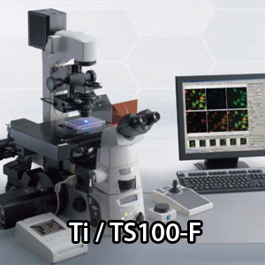
TS100
A new standard for inverted microscopes defined by bright high-resolution images and unrivaled usability
The compact high-performance inverted microscopes ECLIPSE TS100 and TS100-F use Eco-illumination, a newly developed LED illumination. Eco-illumination provides enough brightness for phase contrast and NAMC observations. With a fly-eye lens, uniform brightness is provided in the entire field of view. LEDs are an environmentally friendly low-power-consumption light source. Eco-illumination provides a long lifetime of 60,000 hours and reduces the frequency of lamp replacement. A halogen illumination model is also available.
Nikon's highly acclaimed CFI60 optical system is used, providing flat, sharp and clear images, while achieving longer working distances and higher numerical apertures.
Simple, strain-free operation
The coaxial coarse/fine focus knob located in front of and close to the operator makes operation at high magnifications easier than ever before.
Efficient, user-friendly stage
The stage features a low-profile design that is 195 mm high, making it the ideal size for a lab bench or safety hood. Even cell cultures on the bottom of a tall flask or stacking chamber vessel can be viewed, because there is 190 mm of space above the stage when the condenser is removed.
Moreover, an acrylic stage ring makes it easy to confirm which objective is being used without removing the specimen from the stage.
Phase contrast microscopy has never been easier, thanks to Nikon's innovative Apodized Phase Contrast objectives
To improve images under this method, Nikon developed an innovative series of Apodized Phase Contrast objectives. These objectives produce images with excellent contrast and a much wider tonal range, shedding light on minute details within a specimen.
Model TS100-F is also available.
To accommodate image documentation, Nikon offers a trinocular model as well. The TS100-F comes with a photo port that accepts various digital camera systems.