|
|
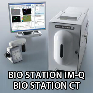
| BIO STATION IM-Q
|
 |
| The perfect solution for long-term, time-lapse, live-cell imaging
|
The BioStation IM-Q incorporates a microscope, an incubator and a high-sensitivity cooled CCD camera in a compact body. This all-in-one package provides a stable environment for live cells and advanced solutions for simple long-term time-lapse data acquisition.
The BioStation IM-Q eliminates the need for a darkroom for fluorescence imaging, meaning it can be installed anywhere.
|
| Exceptional high-sensitivity fluorescence images
|
|
The high sensitivity of the built-in CCD camera, equivalent to ISO800, reduces exposure time, which minimizes photobleaching and damage to specimens while increasing throughput for multipoint acquisition. The cooling mechanism prevents heat-induced noise and allows even weak fluorescence to be captured. The fluorescence equipment employs a noise terminator mechanism in order to eliminate stray light. This enables high-contrast imaging with high S/N ratios.
|
 |
| Cell-friendly environmental control
|
|
Cell culture and image capture functions are beautifully integrated. No complex setup or alignment procedures that conventional time-lapse observation systems require are necessary. The vibration-proof and heat-insulated structure enables stable image acquisition even over long periods.
|
 |
| Accurate recording of live-cell dynamics over a number of days
|
|
Focus drift during lengthy time-lapse observations has been greatly reduced, enabling reliable time-lapse imaging even for several days. The temperature of the chamber, the chamber exterior and microscope unit is precisely controlled with built-in heaters and fans. The focusing mechanism is made from thermal-stable materials and is therefore resistant to thermal expansion. Moreover, BioStation IM-Q moves the objective lens instead of the stage. This eliminates focus drift caused by vibration of culture dish, minimizing stress on the cells.
|
| Lengthy time-lapse imaging without focus drift
|
 |
| Simple and easy operation |
| Operation from a PC |
|
BioStation IM-Q provides fully motorized control from a PC, allowing users who are not accustomed to operating a microscope or camera to easily conduct time-lapse imaging. The user only has to set the culture dish in place and program the image recording.
|
 |
1. Culture dish setup .
|
|
 |
2. Setting with intuitive GUI
1.Set live image location with XYZ movement control.
2.Configure imaging condition.
3.Select illuminations, filters and magnifications.
4.Input imaging time interval.
5.Start time-lapse imaging. . .
|
|
 |
3. Time-lapse imaging and analysis . .
|
|
| Image file output at desired Z-axis planes |
|
Images at different Z-axis planes can be selected from the captured Z-stack images at each time point and assembled into a seamless movie file. This is optimal for imaging a specimen in which the observation point moves along the Z-axis direction, such as with cell division.
|

|
| Streaming function |
|
Rapid motion changes such as cardiomyocyte beats are captured by high-speed 10-fps imaging at user-defined time intervals.
|

|
| Ergo controller |
 |
An ergo controller allows X, Y and Z directional movement with an operational feel similar to a microscope. It also allows changeover of magnifications, fluorescence filters, imaging/observation methods and fluorescence shutter ON/OFF. .
|
|
| Widefield display |
 |
Because a wide 6 mm x 6 mm area can be displayed by image stitching, the point of time-lapse observation can be easily specified from the widefield image. .
|
|
| Two kinds of analysis software are available for intended use. (Option) |
| Imaging software NIS-Elements Ar |
|
Nikon's proprietary imaging software NIS-Elements Ar allows multi-dimensional image capture, image processing, and data management and analysis of up to 6D.
|
| Deconvolution |
 |
Haze and blur of an image that can occur when capturing a thick specimen or a fluorescence image can be eliminated from the captured image.
|
|
| Cell counting |
 |
Images are processed by image analysis routines and the extracted objects can be counted.
|
|
| Slice view |
 |
Images in three orthogonal planes (sliced images along the XY-, YZ-, and ZX-planes) can be viewed in a single display.
|
|
| Volume rendering |
 |
3D images can be reconstructed from captured Z-stack images.
|
|
| Dedicated image analysis software for BioStation series-CL-Quant |
| Cell detection in phase contrast images |
|
CL-Quant automatically detects and measures the cellular area in unstained, label-free phase contrast images. Unique image processing algorithms provide accurate thresholding of phase contrast images, which enables non-invasive quantitative analysis of cells. Cell detection accuracy can be improved through a learning function process.
|

|
| Optional accessories broaden the range of applications
|
| Motorized chamber for four-well culture dish |
|
Automated changeover allows each of the four culture wells to be imaged. Observation of four different samples is possible in a single time-lapse experiment, facilitating comparative analyses.
|

|
| Specialized Hi-Q4 culture dish that enables multi-sample observation |
|
This new 35 mm culture dish is divided into four parts and has an incorporated plane parallel top plate. The plate prevents light path distortion by the meniscus, which is the curve at the air-water interface, and enables high-quality phase contrast observation.
|

|
| Perfusion components |
 |
The perfusion components allow reagent administration and medium exchange without a change to the culture environment. .
|
|
| Two—bottle installation |

|
| BIO STATION CT
|
|
| Stem cell screening inside the incubator |
 |
With conventional cell monitoring procedures, a culture vessel has to be taken out of the incubator for microscope observation, where cells are subjected to stressful environmental changes and vibration. Researchers then have to spend additional time repositioning the vessel to find the same observation points. Nikon's BioStation CT eliminates these problems by providing a stable environment so that the cultures don't suffer while they are being imaged and allowing for a complete trace of the same live cells, including stem cells.
|
|
| Advanced basic functions
|
| Automatic image capture |
 |
The autofocus mechanism allows the capture of in-focus images. Z-stack imaging in phase contrast observation, multi-sample imaging and multi-point imaging are possible with multiple magnifications. User-configured imaging conditions that can be saved in BioStation CT support the repeatability of observations.
|
|
| Automatic vessel transportation |
 |
BioStation CT incorporates a transport unit that provides stable vessel transportation within the heated and humidified incubation area. The high-precision motorized stage in the observation unit allows for automated imaging of the entire area of a well in all culturing formats.
|
|
| Remote access |
 |
Configuring the imaging settings, scheduling a time-lapse experiment, and viewing the cell images are possible via a network. The captured data can be automatically downloaded to the user's local computer. This enables users to monitor the cell status away from the laboratory. When a culture environment (temperature, humidity, CO2 concentration) control error occurs, BioStation CT can notify the users of the error by e-mails.
|
|
| Various functions
|
| Full-well scan imaging and highly magnified image stitching |
|
High-resolution full-well scans are reconstructed by stitching captured adjacent images. This enables clear detection of an iPS colony, which is difficult to detect because of its low induction efficiency, no matter where it forms in the vessel. The specified position of the vessel can be highly magnified with high resolution. BioStation CT also offers cell registration to allow for repeated visits to the same location. These time-lapse sequences can be created even when a vessel is removed from the BioStation CT for medium exchange.
|
| Micro observation |
 |
Phase contrast and fluorescence images can be captured with the high-sensitivity cooled CCD camera. These images can be magnified in 2x, 4x, 10x, 20x and 40x. Up to 40 phase contrast images can be captured along the Z axis with the Z-stack function.
|
|
| Macro observation |
 |
Brightfield image of the whole vessel provides users outside the BioStation CT with information such as handwritten information on the vessel, medium color and whether mold is growing or not. In addition, alkaline phosphatase stained cell counting is possible with the optional image analysis software CL-Quant.
|
|
| Stable culture environment maintenance
|
| Precise temperature control |
 |
The inside temperature is directly controlled by panel heaters embedded in the incubator's six sides. This allows highly precise temperature maintenance. |
|
| Humidity control with air-flow type active aerosol spray humidifier |
 |
Distilled water is automatically sprayed inside the incubator to keep the optimum humidity. Water can be supplied to the tank without opening the incubator door. This air-flow type humidifier reduces contamination risks compared to the water bath type.
|
|
| Environment data recording |
 |
The culture environment is constantly monitored and recorded. The environment data can be accessed at anytime.
|
|
| Smooth vessel transportation |
 |
The waver of liquid surface during the transportation is less than 2 mm. The drift and stress of cells are reduced.
|
|
| Reduced contamination risk
|
|
Rapid motion changes such as cardiomyocyte beats are captured by high-speed 10-fps imaging at user-defined time intervals.
|
| Different GUI for each vessel type
|

|
| Easy operations |
 |
1. Vessel installationM
Culture vessel installation into the storage rack
Efficient installation with the optional sliding storage rack that enables multiple vessels to be installed at one time
|
|
 |
2. Imaging parameter setting
Easy touchscreen operation
Time-lapse imaging configurations such as magnification, imaging point, fluorescence channel and stage motion speed can be set.
|
|
 |
3. Scheduling
Time-lapse imaging schedule
The imaging interval and total period can be set. The shortest time-lapse imaging interval is one minute.
|
|
 |
4. Captured image view
Culture history data management
The time-course change of a specimen can be observed easily in sequentially displayed captured images.
|
|
 |
5. Medium exchange
High-precision repeatability
Accurate tracing of same cells, even after medium exchange, is possible using a dedicated tray holder, as BioStation CT records culture history, such as medium exchange and subculture, as well as X-Y positions for each vessel.
|
|
 |
6. Data report
Reliable data management and documentation support
Obtained data is duplicated and protected using uninterruptible power supply.
Observation information such as temperature, humidity and imaging date can be written and displayed on the captured image to simplify presentation document preparation.
|
|