These microscopes enables educidation of structure and function of the nano-Scopic machinery within living cells.
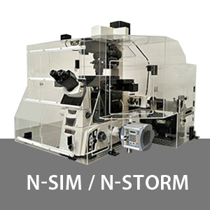
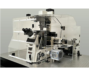
N-SIM
Temporal resolution of 0.6 sec./frame enables super resolution time-lapse imaging of dynamic live cell events
In Structured Illumination Microscopy, the unknown cellular ultra-structure is elucidated by analyzing the moiré pattern produced when illuminating the specimen with a known high-frequency patterned illumination. Nikon's Structured Illumination Microscopy (N-SIM) realizes super resolution of up to 85nm in multiple colors. In addition, it can continuously capture super resolution images at temporal resolution of 0.6 sec./frame, enabling the study of dynamic interactions in living cells. quality and flexibility.
Live cell imaging at double (to approx. 85nm) the resolution of conventional optical microscope
The N-SIM super resolution microscope utilizes Nikon's innovative new approach to "Structured Illumination Microscopy" technology. By pairing this powerful technology with Nikon's renowned CFI Apo TIRF 100x oil objective lens (NA 1.49), N-SIM nearly doubles (to approx. 85nm*) the spatial resolution of conventional optical microscopes, and enables detailed visualization of the minute intracellular structures and their interactive functions.
*Excited with 488nm laser, in TIRF-SIM mode
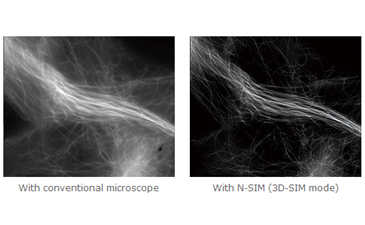
Microtubules in B16 melanoma cell labeled with YFP
Objective: CFI Apo TIRF 100x oil (NA 1.49) Image capturing speed: approx. 1.8 sec./frame (movie)
Photographed with the cooperation of: Yasushi Okada, Ph.D., Department of Cell Biology and Anatomy, Graduate School of Medicine, University of Tokyo
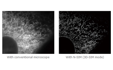
Endoplasmic reticulum (ER) in living HeLa cell labeled with GFP
Objective: CFI Apo TIRF 100x oil (NA 1.49) Image capturing speed: approx. 1.5 sec./frame (movie)
Photographed with the cooperation of: Ikuo Wada, Ph.D., Institute of Biomedical Sciences, Fukushima Medical University School of Medicine
Temporal resolution of 0.6 sec./frame–amazingly fast super resolution microscope system
N-SIM provides ultra fast imaging capability for Structured Illumination techniques, with a time resolution of up to 0.6 sec/frame, which is effective for live-cell imaging (with TIRF-SIM/2D-SIM mode; imaging of up to approx. 1 sec./frame is possible with 3D-SIM mode).
Various observation modes
TIRF-SIM/2D-SIM mode
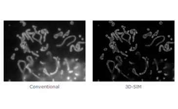
This mode captures super resolution 2D images at high speed with incredible contrast. TIRF-SIM takes advantage of Total Internal Reflection Fluorescence observation at double the resolution as compared to conventional TIRF microscopes, facilitating a greater understanding of molecular interactions at the cell surface.
Plasma membrane of B16 melanoma cell labeled with YFP
* Objective: CFI Apo TIRF 100x oil (NA 1.49)
* Photographed with the cooperation of: Yasushi Okada, Ph.D., Department of Cell Biology and Anatomy, Graduate School of Medicine, University of Tokyo
3D SIM capable
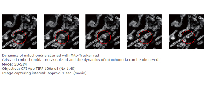
TAxial super resolution observation using the N-SIM system enables optical sectioning of specimens at 300nm resolution in cells and tissues of up to 20µm thickness. Additionally 3D SIM eliminates out of focus background fluorescence resulting in breathtaking contrast.
Mitochondria of NIH-3T3 cell labeling MitoTrackerRed. The Crista is observed clearly by N-SIM
5 laser multi-color super resolution capability
The Nikon LU-5 is a modular system with up to 5 lasers enabling true multi-spectral super resolution. Multi-spectral capability is essential to the study of dynamic interactions of multiple proteins of interest at the molecular level.
*Excited with 488nm laser, in TIRF-SIM mode
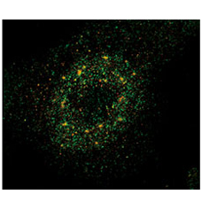
Co-localization images of a target protein of VGEF signaling (Cy3) and its ubiquitin E3 ligase (FITC)
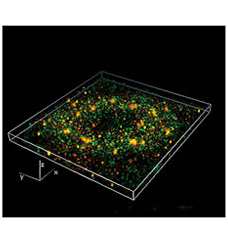
Unprecedentedly detailed structure of the nuclear body can be observed
Mode: 3D-SIM, Z-stack
Objective: CFI Apo TIRF 100x oil (NA 1.49)
Photographed with the cooperation of: Hidetaka Ohnuki, Ph.D., Shigeki Higashiyama, Ph.D., Ehime University Graduate School of Medicine
3D reconstruction image approx. 5µm thick (part)
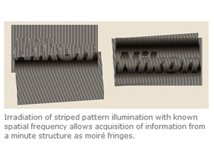
The Principle of the Structured Illumination Microscopy
Analytical processing of recorded moiré patterns produced by overlay of a known high spatial frequency pattern mathematically restores sub-resolution structure of a specimen.
Utilization of high spatial frequency laser interference to illuminate sub-resolution structure within a specimen produces moiré fringes, which are captured. These moiré fringes include modulated information of the sub-resolution structure of the specimen.
Through image processing, the unknown specimen information can be recovered to achieve resolution beyond the limit of conventional optical microscopes.
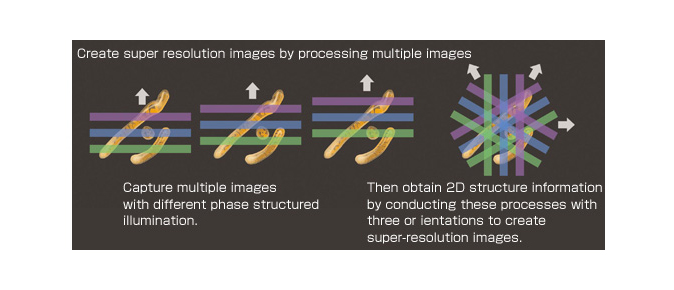
Create super resolution images by processing multiple moiré pattern images
An image of moiré patterns captured in this process includes information of the minute structures within a specimen. Multiple phases and orientations of structured illumination are captured, and the displaced "super resolution" information is extracted from moiré fringe information. This information is combined mathematically in "Fourier" or aperture space then transformed back into image space creating an image at double the conventional resolution limit.
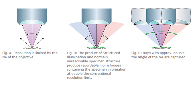
Utilizing High Frequency striped illumination to double the resolution
The capture of high resolution, high spatial frequency information is limited by the Numerical Aperture (NA) of the objectives, and spatial frequencies of structure beyond the optical system aperture are excluded (Fig. A). Illuminating the specimen with high frequency structured illumination, which is multiplied by the unknown structure in the specimen beyond the classical resolution limit, brings the displaced "super resolution" information within the optical system aperture (Fig. B).
When this "super resolution" information is then mathematically combined with the standard information captured by the objective lens, it results in an effective doubling of the NA, and therefore resolution of the optical system (Fig. C).
N-STORM
Achieving a resolution 10 times greater than a conventional optical microscope enables molecular level understanding
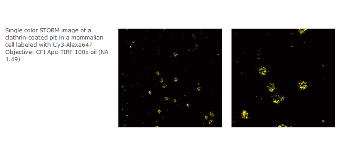
STochastic Optical Reconstruction Microscopy (STORM) reconstructs a super resolution fluorescent image by combining precisely localized information of each fluorophore detected within a complex microscope specimen. N-STORM applies high-accuracy multi-channel molecular localization and reconstruction in 3 dimensions taking full advantage of Nikon's powerful Ti-E inverted microscope, realizing super resolution of 10 times (approx. 20nm laterally) greater than conventional microscopes. This powerful technology can bring to view nanoscopic molecular interactions opening new worlds of understanding.
Super resolution at 10 times (approx. 20nm laterally) greater than conventional optical microscopes
N-STORM utilizes highly accurate localization information (2D or 3D) of 1000's of discrete fluorophor moleculess within a microscope specimen to create breathtaking "super-resolution" images, exhibiting spatial resolution 10 times greater than conventional optical microscopes.
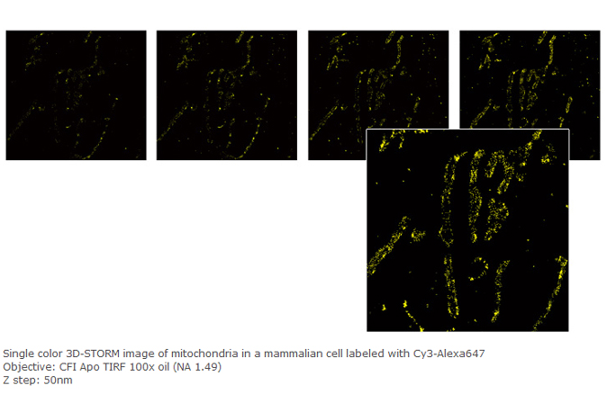
N-STORM can uniquely generate greater than 10 times standard optical resolution axially as well (approx. 50nm)
In addition to lateral super-resolution, N-STORM utilizes proprietary methods to achieve a 10 fold enhancement in axial resolution, effectively providing 3D information at a nanoscopic scale.
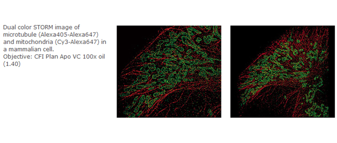
Multi-color imaging using various fluorescent probes
Multi-color super resolution imaging is possible by cleverly combining various "activator" and "reporter" probes. This makes it possible to gain critical insight into the co-localization and interaction of multiple proteins at the molecular level.
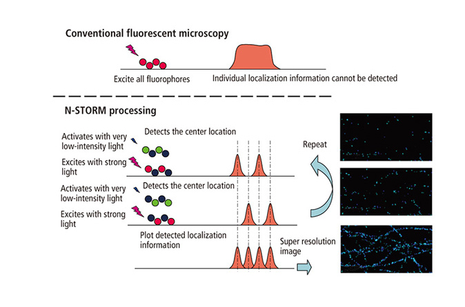
The Principle of N-STORM (STochastic Optical Reconstruction Microscopy)
STochastic Optical Reconstruction Microscopy (STORM) reconstructs a super resolution image by combining the high-accuracy localization information of each fluorophore in 3 spatial dimensions and multiple colors
N-STORM uses stochastic activation of relatively small numbers of fluorophor molecules using very low-intensity light. This low-level stochastic "activation" of discrete molecules enables high precision Gaussian fitting of each laterally. Additionally, taking advantage of an induced astigmatism via the special 3D-STORM optics, N-STORM localizes each molecule axially.
Computationally combining molecular coordinates in 3 dimensions results in high contrast 3D images of the nanoscopic world with molecular specificity.
Dedicated fluorescent dyes
N-STORM uses dedicated fluorescent dye pairs containing an "activator" (relatively short wavelength excitation) and a "reporter" (relatively long wavelength excitation), which enables various color combinations, facilitating true multi-channel super resolution.